Abdominal Anatomy : Abdominal Anatomy. The major organs of the abdomen include the small intestine, large intestine, and stomach. Stomach is a muscular bag forming the most distensible part of the human digestive system. It extends to the lumbar spine, which joins the thorax and pelvis and is a point of attachment for some abdominal wall structures 1 . The rectus abdominis connects to the xiphoid process, a bony landmark at the bottom of the sternum. Top is always the skin side
It is the long, flat muscle that extends vertically between the pubis and the fifth, sixth, and seventh ribs. The human abdomen is divided into quadrants and regions by anatomists and physicians for the purposes of study, diagnosis, and treatment. The major organs of the abdomen include the small intestine, large intestine, and stomach. The abdomen is the part of the body that contains all of the structures between the thorax (chest) and the pelvis, and is separated from the thorax via the diaphragm. Related posts of anatomy of the abdomen women anatomy of the female reproductive system.
Anatomy Of The Abdomen And Pelvis A Journey From Basis To Clinic Coursera from d3njjcbhbojbot.cloudfront.net As its name suggests, the direction of its fibers are obliquely oriented, perpendicular to those of the external abdominal oblique. The division into four quadrants allows the localisation of pain and tenderness, scars, lumps, and other items of interest, narrowing in on which organs and tissues may be involved. It is the long, flat muscle that extends vertically between the pubis and the fifth, sixth, and seventh ribs. It is the most complete reference of human anatomy available on web, ipad, iphone and android devices. The region occupied by the abdomen is called the abdominal cavity, and is enclosed by the abdominal muscles at front and to the sides, and by part of the vertebral column at the back. These organs are held together loosely by connecting tissues. The quadrants are referred to as the left lower quadrant, left upper. Connective tissue called the mesentery holds the abdominal organs together.
It also contains the spleen.
Abdomen, in human anatomy, the body cavity lying between the chest or thorax above and the pelvis below and from the spine in the back to the wall of abdominal muscles in the front. The abdominal wall encloses the abdominal cavity, which holds the bulk of the gastrointestinal viscera. As a general rule, each organ and abnormality is imaged in two directions; By convention, the abdominal exam is performed with the provider standing on the patient's right side. In most cases the transversal and sagittal directions. Top is always the skin side The area occupied by the abdomen is called the abdominal cavity. The abdomen is the part of the body that contains all of the structures between the thorax (chest) and the pelvis, and is separated from the thorax via the diaphragm. Connective tissue called the mesentery holds the abdominal organs together. Topical anatomy of the abdomen. This requires complete exposure of the region in question, which is accomplished as follows: Together, these three turn nutrients into usable energy, as well as help dispose of solid waste. Ct, mri, radiographs, anatomic diagrams and nuclear images.
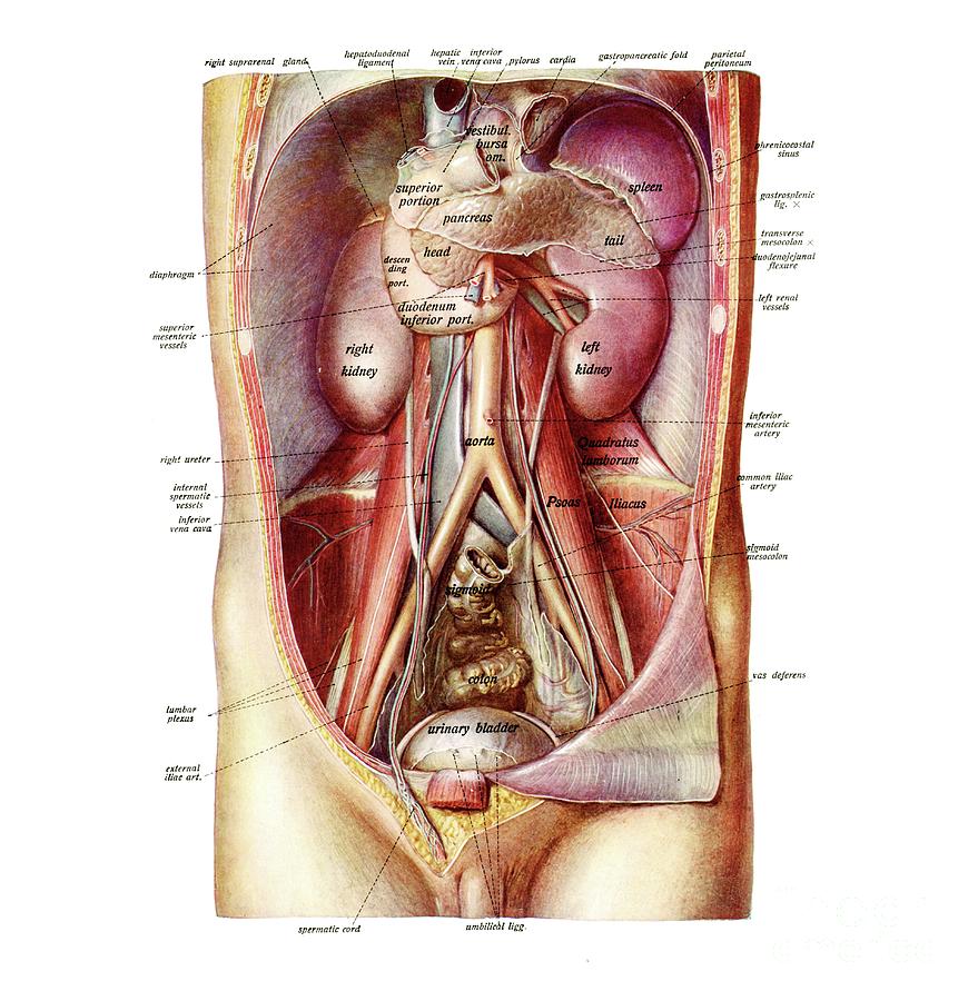
The abdomen contains all the digestive organs, including the stomach, small and large intestines, pancreas, liver, and gallbladder. The major organs of the abdomen include the small intestine, large intestine, and stomach. The anatomy of the regions and planes of the abdomen is composed of many layers with varying blood supply and innervation. Abdominal anatomy includes a major element of the gastrointestinal, system, the caudal end of the oesophagus, stomach, large and small intestine, liver, pancreas and the gallbladder. Browse 7,542 abdomen anatomy stock photos and images available, or search for digestive system to find more great stock photos and pictures.
Anatomy Of Abdomen Photograph By Microscape Science Photo Library from images.fineartamerica.com It extends to the lumbar spine, which joins the thorax and pelvis and is a point of attachment for some abdominal wall structures 1 . The abdominal cavity is the part of the body that houses the stomach, liver, pancreas, kidneys, gallbladder, spleen, and the large and small intestines.the diaphragm marks the top of the abdomen and the horizontal line at the level of the top of the pelvis marks the bottom. Abdominal anatomy includes a major element of the gastrointestinal, system, the caudal end of the oesophagus, stomach, large and small intestine, liver, pancreas and the gallbladder. It is an artery, meaning that it carries blood away from the heart. The layers of the abdominal wall consist of the skin, superficial fascia, and muscles. Explore over 6700 anatomic structures and more than 670 000 translated medical labels. The majority of these organs are encased in a protective membrane termed the peritoneum. The abdomen is the front part of the abdominal segment of the trunk.
The abdomen is the part of the body that contains all of the structures between the thorax (chest) and the pelvis, and is separated from the thorax via the diaphragm.
It is the long, flat muscle that extends vertically between the pubis and the fifth, sixth, and seventh ribs. Tips for viewing stored ultrasound images: The division into four quadrants allows the localisation of pain and tenderness, scars, lumps, and other items of interest, narrowing in on which organs and tissues may be involved. The human abdomen is divided into quadrants and regions by anatomists and physicians for the purposes of study, diagnosis, and treatment. The diaphragm is its upper boundary. Explore over 6700 anatomic structures and more than 670 000 translated medical labels. The rectus abdominis connects to the xiphoid process, a bony landmark at the bottom of the sternum. The quadrants are referred to as the left lower quadrant, left upper. The area occupied by the abdomen is called the abdominal cavity. The component of the urinary system, kidney and the ureter. The abdominal wall surrounds the abdominal cavity, providing it with flexible coverage and protecting the internal organs from damage. Abdominal anatomy includes a major element of the gastrointestinal, system, the caudal end of the oesophagus, stomach, large and small intestine, liver, pancreas and the gallbladder. The majority of these organs are encased in a protective membrane termed the peritoneum.
The abdomen has been bisected, trisected, and even divided into as many as 9 separate regions. It extends to the lumbar spine, which joins the thorax and pelvis and is a point of attachment for some abdominal wall structures 1 . In this article, we shall look at the layers of this wall, its surface anatomy and common surgical incisions that can be made to access the abdominal cavity. This requires complete exposure of the region in question, which is accomplished as follows: Abdomen anatomy the abdomen is comprised primarily of the digestive tract and other accessory organs which assist in digestion, the urinary system, spleen, and the abdominal muscles (shown below).
Male Internal Anatomy Of Chest And Abdominal Area On Black Background Stocktrek Images from www.stocktrekimages.com Ct, mri, radiographs, anatomic diagrams and nuclear images. Together, these three turn nutrients into usable energy, as well as help dispose of solid waste. The abdominal wall is defined cranially by the xiphoid process of the sternum and the costal margins, and caudally by the iliac and pubic bones of the pelvis. The diaphragm is its upper boundary. Together, these three turn nutrients into usable energy, as well as help dispose of solid waste. In most cases the transversal and sagittal directions. The human abdomen is divided into quadrants and regions by anatomists and physicians for the purposes of study, diagnosis, and treatment. Related posts of anatomy of the abdomen women anatomy of the female reproductive system.
In anatomy and physiology, you'll learn how to divide the abdomen into nine different regions and four different quadrants.
Abdomen, in human anatomy, the body cavity lying between the chest or thorax above and the pelvis below and from the spine in the back to the wall of abdominal muscles in the front. Tips for viewing stored ultrasound images: It extends to the lumbar spine, which joins the thorax and pelvis and is a point of attachment for some abdominal wall structures 1 . This requires complete exposure of the region in question, which is accomplished as follows: The abdomen is the front part of the abdominal segment of the trunk. Browse 7,542 abdomen anatomy stock photos and images available, or search for digestive system to find more great stock photos and pictures. Related posts of anatomy of the abdomen women anatomy of the female reproductive system. The abdominal aorta enters the abdomen through the diaphragm at the level of the twelfth thoracic vertebre and continues to just below the umbilical area, where it splits into the right and left common iliac arteries. The area occupied by the abdomen is called the abdominal cavity. These organs are held together loosely by connecting tissues. As its name suggests, the direction of its fibers are obliquely oriented, perpendicular to those of the external abdominal oblique. The rectus abdominis connects to the xiphoid process, a bony landmark at the bottom of the sternum. If you plan to enter a healthcare profession such as nursing, this is something you'll use on the job when performing abdominal assessments (and while documenting).
Share :
Post a Comment
for "Abdominal Anatomy : Abdominal Anatomy"


Post a Comment for "Abdominal Anatomy : Abdominal Anatomy"